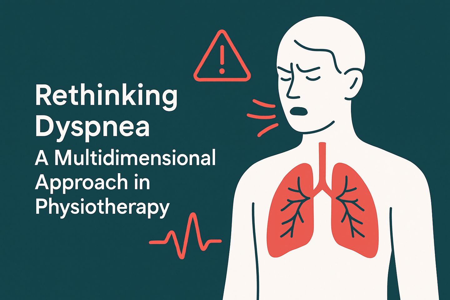
Dyspnea: A Multidimensional Approach in Physiotherapy
STUDY MATERIAL
PhysioAlchemy
7/29/2025
Introduction
Dyspnea, commonly referred to as shortness of breath, is a complex, multidimensional experience that affects individuals across a wide range of cardiopulmonary, neurological, and musculoskeletal conditions. Despite its high prevalence and significant impact on functional status and quality of life, dyspnea is often assessed and managed inadequately within clinical physiotherapy practice.
Traditional clinical models have predominantly approached dyspnea from a mechanistic standpoint, focusing primarily on gas exchange abnormalities, ventilatory inefficiency, and respiratory mechanics. However, emerging literature emphasizes that dyspnea is not solely a physiological response but a subjective experience influenced by sensory input, affective perception, cognitive processing, and behavioral response.
In light of this understanding, it becomes imperative for physiotherapists to adopt a multidimensional framework for the assessment and management of dyspnea. This article aims to provide a comprehensive overview of the underlying mechanisms, clinical assessment tools, and evidence-based physiotherapeutic interventions aligned with current research and multidimensional models of dyspnea.
Understanding Dyspnea: More Than a Symptom
Dyspnea is defined by the American Thoracic Society (ATS) as a "subjective experience of breathing discomfort that consists of qualitatively distinct sensations that vary in intensity" (Parshall et al., 2012). It is a multidimensional construct encompassing physiological, psychological, environmental, and social components.
Dyspnea arises due to a mismatch between respiratory demand and the ability of the respiratory system to meet that demand. This includes both actual and perceived imbalance in the effort to breathe.
Increased Respiratory Drive:
Chemoreceptor activation (central and peripheral) in response to hypoxemia, hypercapnia, or acidosis stimulates the respiratory centers in the brainstem.
The brain interprets this increased neural input as a need to breathe more, leading to a subjective sense of breathlessness.
Impaired Pulmonary Mechanics:
Conditions such as asthma, COPD, interstitial lung disease, or pleural effusion lead to airflow obstruction, reduced lung compliance, or restriction of lung expansion.
These mechanical limitations increase the work of breathing, contributing to the sensation of dyspnea.
Respiratory Muscle Dysfunction:
Fatigue, neuromuscular diseases, or mechanical disadvantage (e.g., hyperinflation in COPD) impairs the efficiency of respiratory muscles.
The patient perceives more effort is required to achieve ventilation, exacerbating dyspnea.
Ventilation-Perfusion (V/Q) Mismatch:
Inefficient gas exchange due to conditions like pulmonary embolism or pneumonia causes hypoxemia.
The body increases the respiratory rate to compensate, which may not be sufficient, resulting in persistent breathlessness.
Cardiovascular Contribution:
In heart failure, increased pulmonary capillary pressures cause interstitial and alveolar edema.
This stiffens the lungs and stimulates pulmonary J receptors, creating a sensation of air hunger or unsatisfied inspiration.
Abnormal Central Perception:
The brain integrates signals from chemoreceptors, mechanoreceptors, and cortical inputs.
In conditions like anxiety, panic disorder, or after chronic disease adaptation, this integration may be exaggerated, leading to a heightened perception of dyspnea even without significant physical abnormalities.
Reduced Respiratory Reserve:
Individuals with compromised lung function have less buffer capacity during exertion.
Even mild physical activity can provoke a respiratory demand that exceeds their reserve, triggering dyspnea.
Dynamic Hyperinflation (in COPD):
Trapped air during expiration leads to an increase in end-expiratory lung volume.
This reduces inspiratory capacity, creates mechanical disadvantage for the diaphragm, and intensifies the sensation of breathlessness.
Role of Psychological Factors:
Anxiety and emotional distress amplify the perception of breathlessness.
The limbic system and cortical awareness can magnify respiratory sensations, leading to disproportionate subjective symptoms compared to objective findings.
Assessment: From Numbers to Nuance
In clinical practice, dyspnea is often assessed using simple, unidimensional tools like the Modified Borg Scale, Visual Analog Scale (VAS), or the Medical Research Council (MRC) Dyspnea Scale. These scales serve a purpose.
A. Subjective Assessment Tools (Patient-Reported Scales)
These tools quantify the patient’s perception of breathlessness.
Modified Borg Dyspnea Scale: Rates perceived breathlessness from 0 (no breathlessness) to 10 (maximal breathlessness). It is often used during exercise testing (e.g., 6MWT, CPET).
Visual Analog Scale (VAS): A 10 cm horizontal line where the patient marks their dyspnea level. Left end: No dyspnea, Right end: Worst imaginable dyspnea.
Medical Research Council (MRC) Dyspnea Scale / Modified MRC (mMRC): Grades 0–4 describing dyspnea severity based on activity limitation. It is widely used in chronic respiratory diseases, especially COPD.
Baseline Dyspnea Index (BDI) and Transition Dyspnea Index (TDI): BDI measures initial dyspnea severity in three domains: Functional impairment, Magnitude of task, Magnitude of effort. TDI tracks changes over time, useful in monitoring treatment outcomes.
Dyspnea-12 Questionnaire: Assesses both physical and emotional components of breathlessness. It contains 12 descriptors rated on a 0–3 scale, total score 0–36.
B. Objective Assessment Tools (Physiological and Functional Measures)
Pulse Oximetry: Measures oxygen saturation (SpO₂); helps correlate desaturation with dyspnea. It is important in monitoring hypoxemia during exertion.
Arterial Blood Gases (ABG): It measures PaO₂, PaCO₂, pH, and HCO₃. It helps identify respiratory/metabolic imbalances associated with dyspnea.
Pulmonary Function Tests (PFTs): It evaluates the lung volumes, capacities, flow rates. The common parameters includes: FEV₁, FVC, FEV₁/FVC ratio. It helps to distinguish between obstructive vs. restrictive lung diseases.
6-Minute Walk Test (6MWT): It assesses functional exercise capacity and dyspnea on exertion. Dyspnea scores often recorded using Borg Scale before and after test.
Cardiopulmonary Exercise Testing (CPET): It measures VO₂ max, ventilatory threshold, and dyspnea response. The gold standard for assessing integrated cardiopulmonary function.
Chest Imaging (X-ray / CT scan): It detects anatomical or pathological causes of dyspnea (e.g., fibrosis, effusion, collapse). It is especially important when symptoms are disproportionate to functional tests.
C. Multidimensional Assessment
Dyspnea is not purely physiological; it has emotional, functional, and cognitive dimensions.
Multidimensional tools help in holistic evaluation and management: (i) Combine patient self-report, objective measures, and functional status, and (ii) Useful in palliative care, chronic disease management, and rehabilitation settings.
Clinical Presentations of Dyspnea Across Systems
Dyspnea manifests across a broad spectrum of pathologies, with varying underlying mechanisms depending on the system involved. A system-specific approach enables more precise clinical reasoning and targeted physiotherapeutic intervention.
Pulmonary System
In pulmonary conditions such as chronic obstructive pulmonary disease (COPD), interstitial lung disease (ILD), and asthma, dyspnea typically arises due to airway obstruction, parenchymal restriction, impaired gas exchange, and dynamic hyperinflation. Patients may describe sensations such as "air hunger" or "increased work of breathing."
Cardiovascular System
In cardiovascular conditions such as congestive heart failure (CHF) or ischemic heart disease, dyspnea is commonly associated with reduced cardiac output, increased pulmonary capillary pressure, and fluid retention. It often presents as exertional dyspnea, orthopnea, or paroxysmal nocturnal dyspnea.
Neurological Conditions
Neurological disorders including Parkinson’s disease, stroke, motor neuron disease, and spinal cord injury may present with dyspnea secondary to impaired central respiratory drive, neuromuscular weakness, or altered respiratory coordination.
Musculoskeletal and Postural Disorders
Structural abnormalities such as severe kyphoscoliosis, ankylosing spondylitis, or post-operative thoracic stiffness can mechanically restrict thoracic expansion and diaphragmatic excursion, resulting in perceived breathlessness.
Post-COVID and Long COVID Syndromes
Dyspnea is a persistent and disabling symptom in many patients recovering from COVID-19, often out of proportion to imaging or pulmonary function test results. Mechanisms may include autonomic dysfunction, deconditioning, breathing pattern disorders, and anxiety.
Physiotherapy Intervention for Dyspnea
The primary goal of physiotherapy in patients experiencing dyspnea is to reduce the sensation of breathlessness, improve ventilatory mechanics, optimize oxygen delivery, enhance functional capacity, and improve quality of life. Physiotherapists use evidence-based interventions targeting respiratory muscle function, airway clearance, breathing patterns, physical conditioning, and anxiety associated with breathlessness. Interventions are individualized based on the underlying pathology, severity of dyspnea, and patient-specific goals.
Breathing Retraining
Breathing retraining focuses on correcting abnormal or inefficient breathing patterns commonly observed in dyspneic individuals.
Diaphragmatic Breathing: This technique promotes abdominal expansion during inhalation, thereby improving diaphragmatic excursion, reducing accessory muscle use, and lowering the work of breathing. It is particularly beneficial in conditions like chronic obstructive pulmonary disease (COPD) and restrictive lung disorders.
Pursed-Lip Breathing (PLB): Widely used in obstructive pulmonary conditions, PLB helps in delaying airway collapse during exhalation, maintaining positive airway pressure, and reducing dynamic hyperinflation.
Segmental Breathing Exercises: These target specific lung segments to enhance localized ventilation, useful postoperatively or in cases of pleural pathology.
Positioning Strategies
Body positioning significantly influences ventilation-perfusion (V/Q) matching and respiratory muscle efficiency.
High Fowler's Position (45–90° upright) and Forward Lean Sitting (Tripod Position) help in optimizing diaphragmatic mechanics and reducing dyspnea by facilitating accessory muscle activation.
Side-lying with the affected lung uppermost may enhance oxygenation by improving perfusion to the less-affected lung.
Gravity-Assisted Drainage Positions may also be employed when airway clearance is needed in addition to dyspnea relief.
Respiratory Muscle Training (RMT)
RMT involves the use of resistive devices or threshold-loading trainers to improve the strength and endurance of respiratory musculature, especially the diaphragm and accessory muscles.
Inspiratory Muscle Training (IMT) is commonly prescribed using threshold or pressure-loading devices. Studies have shown significant improvements in inspiratory muscle strength, dyspnea scores, and exercise capacity in COPD, congestive heart failure, and neuromuscular conditions.
Expiratory Muscle Training (EMT) may be beneficial in patients with ineffective cough or impaired airway clearance.
Airway Clearance Techniques (ACTs)
In patients where dyspnea is aggravated by retained secretions, ACTs play a crucial role.
Active Cycle of Breathing Technique (ACBT), Autogenic Drainage, Manual Chest Physiotherapy, and Positive Expiratory Pressure (PEP) therapy are used to mobilize and clear mucus, reducing airflow obstruction and dyspnea.
Mechanical devices like high-frequency chest wall oscillation or mechanical insufflation–exsufflation may be indicated in patients with neuromuscular weakness.
Physical Reconditioning and Exercise Training
Exercise intolerance due to dyspnea often leads to deconditioning, which in turn worsens breathlessness a vicious cycle that physiotherapy aims to break.
Aerobic Training (e.g., treadmill walking, cycling, arm ergometry) improves cardiovascular endurance and reduces dyspnea on exertion. The intensity is usually guided by symptom-limited tests or using the Borg Dyspnea Scale.
Strength Training for both upper and lower limbs is incorporated to enhance overall functional capacity and reduce oxygen consumption during daily tasks.
Interval Training may be more tolerable for patients with severe breathlessness.
Pulmonary rehabilitation programs include multidisciplinary exercise protocols and education, demonstrating strong evidence in reducing dyspnea, especially in chronic lung diseases.
Energy Conservation Techniques
Physiotherapists educate patients in energy-saving methods for daily tasks:
Breaking tasks into smaller steps
Using assistive devices
Avoiding unnecessary movements
Synchronizing breathing with effort
These strategies help in reducing the oxygen demand of routine activities.
Relaxation and Psychosocial Support
Since anxiety and panic can exacerbate dyspnea, relaxation techniques such as:
Progressive Muscle Relaxation (PMR)
Guided imagery
Mindfulness-based breathing exercises
are incorporated into rehabilitation programs. Physiotherapists also collaborate with psychologists when emotional factors significantly contribute to dyspnea.
Use of Adjunctive Modalities
Fan Therapy: Directing cool air over the face (especially around the trigeminal area) has been found to reduce the sensation of breathlessness through neurosensory mechanisms.
TENS (Transcutaneous Electrical Nerve Stimulation) applied to thoracic areas has been studied as a non-pharmacological method to manage dyspnea, especially in palliative care.
Supplemental Oxygen: While not directly under physiotherapy scope, its appropriate use in hypoxemic patients enhances the effectiveness of rehabilitation and exercise tolerance.
Case-Based Questions: Dyspnea
A 65-year-old male patient with a 40-pack-year smoking history presents with acute breathlessness, wheezing, and productive cough for the past 3 days. His respiratory rate is 28/min, SpO₂ is 88% on room air. He has a past diagnosis of COPD.
Q1: What is the most likely cause of dyspnea in this patient?
Q2: What clinical features will you assess as a physiotherapist?
Q3: List 3 physiotherapy goals for this patient during acute exacerbation.
Q4: Name and justify any two physiotherapy techniques you would use to manage dyspnea in this case.
A 58-year-old female is 4 days post-CABG. She complains of breathlessness while walking and performing bed mobility tasks. She has an FEV₁/FVC of 65% and chest X-ray shows basal atelectasis.
Q1: Explain the physiological reason for her dyspnea.
Q2: What objective measures can be used to evaluate dyspnea in this case?
Q3: Which breathing exercises are indicated in the early post-operative period?
Q4: How will you progress ambulation for this patient?
A 40-year-old male diagnosed with Guillain-Barré Syndrome (GBS) presents with increasing difficulty in breathing, weak cough, and SpO₂ of 92% on 2L O₂ via nasal cannula. His MIP (Maximum Inspiratory Pressure) is -20 cmH₂O.
Q1: What is the mechanism of dyspnea in this case?
Q2: Which physiotherapy assessments are essential for this patient’s respiratory function?
Q3: What are the indications for non-invasive ventilation in this case?
Q4: Describe a physiotherapy intervention to improve cough efficacy.
A 45-year-old male reports persistent dyspnea 6 weeks post mild COVID-19 infection. His CT chest shows fibrotic bands, 6-minute walk distance is 280 meters, and mMRC score is 2.
Q1: What might be the underlying cause of dyspnea in this patient?
Q2: Which exercise testing protocols can be used to monitor progress?
Q3: What physiotherapy components should be included in his pulmonary rehab plan?
Q4: How would you educate the patient about pacing and energy conservation?